Associate Professor Payal Saxena
  • About
  • Areas of Expertise
    • Articles
    • Video Interviews
  • Patient Info
    • UPTODATE
  • Procedures
    • Gastroscopy
    • Colonoscopy
    • Endoscopic Ultrasound (EUS)
    • ERCP
    • Pancreatic Cancer Screening
  • POEM
  • Contact
  • Professional Activities
    • Current
    • Professional appointments & memberships
    • Editorial activities
    • Invited presentations
    • Research & publications

4-step approach to POEM

9/6/2015

0 Comments

 
Payal Saxena, MD, from the Department of Medicine, Division of Gastroenterology and Hepatology at Johns Hopkins Hospital in Baltimore, Maryland presents this video case titled “Peroral endoscopic myotomy: a 4-step approach to a challenging procedure” from the VideoGIE section
Our video demonstrates the techniques for performing each of the 4 steps of POEM, which are (1) mucosal entry, (2) submucosal tunneling, (3) myotomy and (4) closure of mucosal entry. The commonly used endoscopic submucosal dissection (ESD) knives (triangle tip and hybrid knife) and methods for performing submucosal injection of dyed saline (such as the jet injection technique) are reviewed. A 6-8cm myotomy (4-6cm esophageal and 2-3cm gastric) is adequate for most cases. However, a longer myotomy is needed in patients with spastic esophageal disorders. The optimal length can be determined by the proximal extent of spastic contractions on high resolution manometry.

Both selective inner circular and full-thickness myotomies are commonly performed and have been shown to be equally effective. However, selective inner circular myotomy may ensure a safety margin away from mediastinal and peritoneal structures as well as minimizing extraluminal leakage of carbon dioxide. Maintaining integrity of the mucosal flap and reliable closure of the mucosal entry is paramount to ensuring safety of the procedure and preventing perforation and leakage of esophageal contents into the mediastinal space. Standard hemostatic clips are commonly used. Alternate methods of closure include endoscopic suturing or placement of over-the-scope clips. The latter is particularly helpful when closure fails with standard methods.

POEM is a demanding procedure and can be associated with serious adverse events. Therefore, structured training is mandatory. Initially, the endoscopist should observe an experienced operator and be familiar with the all the equipment used for the procedure. Rigorous training in animal models should be performed. Once the endoscopist can consistently perform procedure without complications in an animal mode, clinical procedures are best performed initially under the supervision of an experienced POEM operator.

POEM is a relatively new procedure however increasing numbers are being performed worldwide. We felt an instructive video on the techniques, challenges and methods for training in POEM has great educational benefit for future POEM operators.
0 Comments

EUS-Guided Drainage of Pseudocyst

8/6/2015

0 Comments

 
Payal Saxena, MD, from the Department of Medicine, Division of Gastroenterology and Hepatology at Johns Hopkins Hospital in Baltimore, Maryland, USA presents this video case “EUS-Guided Drainage of a Giant Hemorrhagic Pseudocyst by a Through-the-scope Esophageal Metal Stent.”

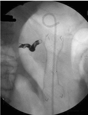
A 78-year-old man with hemorrhagic transformation of a 17cm pseudocyst was hemodynamically unstable requiring splenic artery embolization. He developed gastric outlet obstruction due to the pseudocyst, which was then drained under EUS guidance by creating a cystgastrostomy, flushing the cavity with hydrogen peroxide and placement of a through-the-scope, covered esophageal metal stent (18mm x 60mm) across the cystgastrostomy. A total of 2.4L of blood was drained during the procedure. The patient experienced a rapid resolution of symptoms post-procedure.  At 4 week imaging, the pseudocyst had completely resolved after a single procedure. The stent was easily removed with a snare at follow-up endoscopy.

We have demonstrated a novel technique which facilitates safe and rapid resolution of a giant pseudocyst with large volume solid debris (blood clots) without the need for repeated endoscopic procedures, debridement or external drainage. Hydrogen peroxide facilitated dissolution of the blood clots. The wide bore stent allowed passage of debris from the pseudocyst cavity without becoming clogged. The wide caliber covered metal stent also ensured complete seal of the cystgastrostomy tract, preventing complications of leaks and perforation.

Picture
 Fluoroscopic image of the fully covered self expandable metallic stent placed across the cystgastrostomy. A double pigtail stent is seen within the metallic stent. Splenic artery embolization coil is seen to the left of the stent.
0 Comments

Rendezvous versus direct transluminal techniques for malignant biliary obstruction

8/6/2015

0 Comments

 
Payal Saxena, MD, from Johns Hopkins University in Baltimore, Maryland writes about her Original Article titled “EUS-guided biliary drainage by using a standardized approach for malignant biliary obstruction: rendezvous versus direct transluminal techniques (with videos)” from the November issue. 

The aim of this study was to assess outcomes of EUS-guided biliary drainage (EGBD) performed using a standardized approach in addition to comparing the outcomes of EGBD via the rendezvous technique (REN) or the direct transluminal technique (TL).

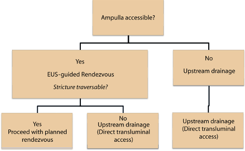
There has been growing international experience with EGBD. The current data suggests that severe adverse events are rare, however most series include a small number of patients. Also, there is no data comparing the REN and TL techniques of EGBD. The REN technique is performed by passing a wire across the papilla and is preferred by many endoscopists as it avoids the need for a permanent bilio-enteric fistual and need for dilatation of the fistulous tract which can lead to adverse events (bleeding, pneumoperitoneum, pneumomediastinum). However, the REN technique is not feasible if the papilla cannot be accessed due to altered anatomy or gastric outlet obstruction. The REN technique has also been associated with a risk of pancreatitis and lengthy procedural times attributed toward wire manipulation. The TL technique can be performed entirely transgastrically or transduodenally without accessing the papilla.

EGBD was performed with technical success in 94% (33/35) of patients, TL technique was used in 20 patients, and REN technique in 13. Clinical success was achieved in 97% (32/33) patients. Only 4 (12%) procedure related adverse events occurred, 2 mild (mild pancreatitis, limited pneumoperitoneum), 1 moderate (retained sheared wire), and 1 severe (acute cholecystitis). There was no statistically significant difference  in clinical success, procedure time, adverse events, or length of hospital stay between the TL and REN groups.

The TL technique has not been widely studied when compared to the REN technique. Our study shows the TL technique is safe and bile leak or pneumoperitoneum can be avoided with the use of metallic stents and carbon dioxide insufflation, respectively. Furthermore, the use of larger covered metallic stents results in complete seal of the iatrogenic bilioenteric tract, preventing bile leak. Our study  demonstrates that use of a standardized approach (Figure 1) allows EGBD to be performed safely and effectively. Prospective, randomized trials are needed to compare the safety and efficacy of the TL and REN techniques of EGBD.

Picture
0 Comments

Antegrade transpapillary placement of a metal biliary stent

8/6/2015

0 Comments

 
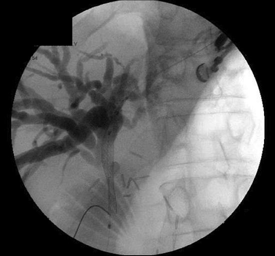
Payal Saxena, MD from Johns Hopkins Hospital in Baltimore, Maryland, USA and the Chris O’Brien Lifehouse at RPA in Camperdown, New South Wales, Australia describes this VideoGIE case “EUS-guided biliary drainage with antegrade transpapillary placement of a metal biliary stent.” EUS-guided biliary drainage with antegrade transpapillary metallic stent is demonstrated in 2cases of malignant, biliary obstruction. Case 1 was a patient with failed ERC due to a long, tight distal CBD stricture. Case 2 was a patient with post-Whipple’s anatomy and recurrence of adenocarcinoma at the hepatojejunostomy. Standard ERC failed due to inability to intubate the afferent limb. In both cases, dilated intrahepatic ducts were identified at EUS. Cholangiogram was performed by injecting contrast after puncture with a 19g needle. A distal stricture (CBD and CHD respectively) was identified in both cases. A 0.025 inch guidewire was passed through the needle and across the stricture followed by balloon dilatation of the stricture with subsequent placement of an uncovered self-expandable metallic stent in an antegrade fashion across the papilla and hepatojejunostomy respectively.

Picture
Figure 1. Cholangiogram obtained via endoscopic ultrasound guided transgastric intrahepatic puncture demonstrates a dilated intrahepatic biliary tree. A self expandable metallic stent is deployed over a wire across a distal common bile duct stricture in an antegrade fashion.
0 Comments
    Picture

    Dr Payal Saxena

    Dr Payal Saxena is a Gastroenterologist and Hepatologist, subspecialised in Interventional Endoscopy. She completed her specialist Gastroenterology training in Sydney then travelled to the USA where she undertook a two-year Interventional Endoscopy and Research fellowship at Johns Hopkins Hospital.


    Categories

    All
    Anastomotic Stenosis


    Archives

    June 2015
    February 2015


    RSS Feed


    Picture
© Dr Payal Saxena 2015
Website by Dalten Media